|
Transcranial
magnetic stimulation (TMS) is a non-invasive technique to stimulate a
restricted part of the cortex. Developed in the 1980s, it has been used
first for clinical diagnostic but rapidly growing interest has shown
potentials for TMS as a therapeutic tool in Psychiatry, as well as an
investigative tool in Cognitive Neuroscience.
How does TMS work?
TMS
is based on the laws of electromagnetic induction: a current passing
through a coil of wire generates a magnetic field perpendicular to the
current direction in the coil. A rapid change of this magnetic field
elicits in turn a transient electric field. This electric field affects
the membrane potential of the nearby neurons, which may lead to
depolarization and neuron discharging or interfere with the ongoing
action potentials. Commercially available stimulators offer the
possibility to generate peak of magnetic field up to 2.5 tesla, with
frequencies up to 30 Hz. Since the magnetic field decreases rapidly
(1/distance 2) only the most external parts of the brain can be targeted with TMS.
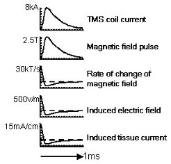
 Time course and order of amplitudes of the events occurring during a TMS pulse. (from Jalinous, 1998, Magstim Company)  Example
of a figure-of-eight TMS coil. Two coils are juxtaposed with the
currents being discharged in opposite directions. The induced electric
fields add up so that the peak of the electric field is located at the
junction between the two coils. (a, b ,c) photograph and in plane and
transversal X-Ray of the coil. (c,d) Induced field two centimetres
above the coil.
What are the applications in cognitive neuroscience?
TMS
has proven a very useful tool to help us understand the functioning of
the normal and pathological human brain. The principal uses of TMS in
cognitive neuroscience include:
1) Probing cortical
excitability by directly measuring the response to single-pulse TMS
applied over primary cortices. In this case the evoked response,
for example, motor evoked potentials for the motor cortex or phosphene
intensity for the visual cortex, as well as the TMS intensity threshold
necessary to elicit those responses can be used as dependent variables. 2)
Interfering briefly with the ongoing cortical activity with single
pulse or short trains of high frequency repetitive TMS. In this
case TMS is applied while the participant is engaged in a behavioural
task. Changes in performance in trials with TMS as compared to trials
without TMS demonstrate the stimulated region was necessary for the
processing involved in the task at the moment of stimulation.
3)
Inducing long-term (minutes) changes in cortical excitability and
connectivity with several minutes of low frequency repetitive TMS. Here
participants participate in behavioural, cognitive or brain imaging
tests before and after the administration of, typically, 10-15 minutes of TMS.
How can the brain area of interest be targeted precisely?
In
order to make inferences about brain-behaviour relationships, it is
crucial to localize precisely the area stimulated by TMS. This is
achieved by identifying this area on the high-resolution magnetic
resonance image (MRI) of the participant. Then, by using frameless
stereotaxy the investigator can position the TMS coil over the scalp so
that the peak of electric field is localized on the desired area.  Frameless stereotaxy Frameless stereotaxy:
In the first step (left), easily identifiable landmarks are marked on
the MRI with a cursor and simultaneously on the head of the subject
with a position tracking device. By using a set of four landmarks,
dedicated software can register the subject’s head to his/her MRI. In
the second step (right), the camera tracks a reflecting device attached
to the coil relatively to the reference, and the software displays the
position of the electric field on the cortical surface. How does TMS complement other methods in cognitive neuroscience?
Transcranial
magnetic stimulation presents the advantage of a precise timing (for
single pulse) and relatively good localisation. The main disadvantage
is the impossibility to stimulate deep brain structures directly. Most
brain imaging techniques allow the investigators to identify brain
areas that are active during a given motor, perceptual or cognitive
process. They cannot tell us, however, whether those areas are necessary for the process. By interfering with the normal functioning of a brain area, TMS enables inferences about a causal link
between this area and behaviour.Second, because of the limited duration
of the interference it induces, TMS can be used to investigate when a brain area is making its critical contribution to behaviour. Thus
TMS offers a very important complement to brain mapping techniques such
as functional MRI or PET. This is illustrated by the growing number of
studies employing a two-step approach: 1) identify brain regions
activated during a given task with brain imaging; 2) target those areas
with TMS to assess and trace the timing of their importance for this
behaviour. In addition, TMS combined directly with PET, fMRI or EEG
allows the researchers to study cortical excitability and connectivity
as well as plasticity, independently of behaviour. While a controlled
stimulation is applied over a given area, brain activity can then be
assessed both locally and in remote brain regions  Combining
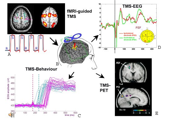
TMS with other methodologies: example of frontal eye field (FEF)
stimulation. A) The FEF can be first localized with fMRI when
alternating periods of saccades (S) with periods of rest (R), either in
individuals or group or with meta-analysis of a corpus of study (e.g.
Grosbras et al Hum Brain Mapp 2005). B) The location identified on the
brain anatomy can then be used as a target for TMS thanks to frameless
stereotaxy. C) TMS applied 50 ms before the expected onset of a saccade
(blue dotted line) delays the saccade. Traces reflect electrooculor
(EOG) activity during auditory cued saccades without TMS (blue) or with
TMS (red). Stimulating a control site show that this effect is specific
to FEF stimulation (Grosbras et al, J Cogn Neurosci,, 2002). D) TMS
applied over the FEF induces electroencephalographic potential as well
as synchronization in parieto-occipital cortices (Grosbras,
unpublished). E) The amount of TMS applied over the FEF is positively
correlated with blood flow locally in the FEF Is TMS safe?
As
functional MRI, TMS is a new investigative technique. Therefore the
scope for appreciating safety is limited to research done in the last
20 years. As for MRI, safety guidelines have to be respected. Within
those guidelines, TMS is considered a safe technique.
Single pulse TMS has
no reported harmful side effects. In some susceptible individuals it
can induce headaches. Those are easily treated with mild analgesic.
Contra-indications for single-pulse TMS are: pace maker, aneurysm clip,
heart/vascular clip, prosthetic valve, intracranial metal prosthesis.
Pregnant women and young children are also excluded from research
studies, although they might be subject to TMS for clinical or
therapeutic purposes. Repetitive TMS
potentially has the power to induce seizures (7 reported cases between
1990 and 1996). Therefore further precautions have to be taken. In
addition to those for single-pulse TMS, exclusion criteria for
repetitive TMS include: personal or familial history of epilepsy,
medications which reduce the threshold for seizure, high alcohol or
drug consumption.
In addition, in an
international workshop held in 1996, safety guidelines have been
established for the parameters of repetitive TMS, which are widely
accepted by the scientific community. Since then no cases of seizure
have been reported.
Selected references
Cowey
A. The Ferrier Lecture 2004 : What can transcranial magnetic
stimulation tell us about how the brain works? Philos Trans R Soc Lond
B Biol Sci. 2005 Jun 29;360(1458):1185-205.
Paus T, Jech R,
Thompson CJ, Comeau R, Peters T, Evans AC. Transcranial magnetic
stimulation during positron emission tomography: a new method for
studying connectivity of the human cerebral cortex. J Neurosci. 1997
May 1;17(9):3178-84.
Virtanen J, Ruohonen J, Naatanen R,
Ilmoniemi RJ. Instrumentation for the measurement of electric brain
responses to transcranial magnetic stimulation. Med Biol Eng Comput.
1999 May;37(3):322-6.
Walsh V, Rushworth M., A primer of magnetic stimulation as a tool for neuropsychology.
Neuropsychologia. 1999 Feb;37(2):125-35. Review.
Wassermann
EM. Risk and safety of repetitive transcranial magnetic stimulation:
report and suggested guidelines from the International Workshop on the
Safety of Repetitive Transcranial Magnetic Stimulation, June 5-7, 1996.
EEG Clin. Neurophysiol. 108:1-16, 1998.
|


 Facilities
  Transcranial Magnetic Stimulation
ęby Marc